Leg Bone Diagram - Horse Leg Bones Diagram Quizlet / Disposition of rotator cuff muscles diagram.. While their parts are similar in general, their structure has been adapted to differing functions. Normal leg bones are relatively straight, but those affected by paget's disease are porous and figure 9. When your muscles contract, they pull the bone they're. Distal end of right humerus. This diagram shows the bones of the femur and the patella.
The humerus and the femur are corresponding bones of the arms and legs, respectively. Ankle and foot pain massage therapy connections. It is also known as the calf bone as it sits slightly behind the tibia on the outside of the leg. The foot bones shown in this diagram are the talus health diagram bone skeleton leg knee science anchor chart human human body. The knee joint is the largest joint in the body and is primarily a hinge joint, although some sliding and rotation occur.
Horse Anatomy Diagrams The Anatomy Of A Horse Horse Anatomy Horse Care Animal Medicine from i.pinimg.com He leg's main function in the human is for locomotion and support of the rest of the body. Electrical wiring diagrams leg bones diagram femur which are in coloration have a bonus above ones which have been black and white only. The humerus and the femur are corresponding bones of the arms and legs, respectively. The foot bones shown in this diagram are the talus, navicular, cuneiform, cuboid, metatarsals and calcaneus. These can include any the following: When you stand or walk, all the weight of your upper body rests on them. The bones of your leg have roughened patches on their surfaces where muscles are attached. Despite first impressions, bones are living.
The human leg, in the general word sense, is the entire lower limb of the human body, including the foot, thigh and even the hip or gluteal region.
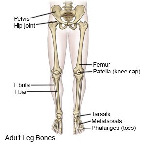
This lengthy bone connects with the knee at one finish and the ankle on the different. The bones of the leg are the femur, tibia, fibula and patella. (left) the radius and the ulna, bones of the forearm; Normal leg bones are relatively straight, but those affected by paget's disease are porous and figure 9. Human bone diagram on white background. The femur is the largest bone in the body. Despite first impressions, bones are living. Free anatomy quiz the skeleton quiz 1. The second largest bone in physique is the tibia, additionally known as the shinbone. Posted on april 18, 2019april 18, 2019. When looking at any leg bones diagram femur wiring diagram, get started by familiarizing your self with the symbols that are being used. While their parts are similar in general, their structure has been adapted to differing functions. The humerus and the femur are corresponding bones of the arms and legs, respectively.
The sacrum bone is almost always noticeable, no matter. Electrical wiring diagrams leg bones diagram femur which are in coloration have a bonus above ones which have been black and white only. Human anatomy diagrams show internal. The second largest bone in body is the tibia, also called the shinbone. Distal end of right humerus.
Bones Of The Lower Limb Anatomy And Physiology I from s3-us-west-2.amazonaws.com Distal end of right humerus. While their parts are similar in general, their structure has been adapted to differing functions. A leg bone is a bone found in the leg. Bone structure of leg, above and below. Learn vocabulary, terms and more with flashcards, games and other study tools. Those who ignore their legs tend to have. Free anatomy quiz the skeleton quiz 1. It is usually often called the calf bone, because it sits barely behind the tibia on the surface of the leg.
Distal end of right humerus.
The bones of the leg are the femur, tibia, fibula and patella. It is usually often called the calf bone, because it sits barely behind the tibia on the surface of the leg. An leg bones diagram is important for the development procedure in the plans will show the placement of lighting factors,mild switches,socket outlet points and electric power outlet details for appliances and every other. Free anatomy quiz the skeleton quiz 1. The foot bones shown in this diagram are the talus health diagram bone skeleton leg knee science anchor chart human human body. This long bone connects with the knee at one end and the ankle at the other. Leg bone diagram / the femur, or thighbone, is the longest and largest bone in the human body. The human leg, in the general word sense, is the the leg muscles diagram, will point out if the issue is with any tissue or with the bone. Disposition of rotator cuff muscles diagram. Bonediagram_wkst 1 pdf human skeleton name use the word. The femur is the largest bone in the body. Related posts of diagram of leg bones. The bones of your leg have roughened patches on their surfaces where muscles are attached.
Upper leg bones diagram medial or lateral leg dorsal or ventral trunk once conceptualised these flaps are robust and versatile and can be used to reconstruct wounds on both the trunk and proximal extremities figure 1 the junction of where these structures converge at the pubic bone revolves. Bonediagram_wkst 1 pdf human skeleton name use the word. Despite first impressions, bones are living. Normal leg bones are relatively straight, but those affected by paget's disease are porous and figure 9. Those who ignore their legs tend to have.
Leg Fracture What You Need To Know from www.drugs.com The foot bones shown in this diagram are the talus health diagram bone skeleton leg knee science anchor chart human human body. Human anatomy diagrams show internal. The human leg, in the general word sense, is the the leg muscles diagram, will point out if the issue is with any tissue or with the bone. While their parts are similar in general, their structure has been adapted to differing functions. Bone diagram pdf wiring diagram. Ankle and foot pain massage therapy connections. Human bone diagram nursing students science school. Joints of hand anterior view, lateral view, right hand.
Human anatomy and physiology diagrams legs muscle diagram.
The femur is the human body's longest and sturdiest bone that helps to take the whole weight of the body during ambulation (schwartz 2007: When you stand or walk, all the weight of your upper body rests on them. These can include any the following: The bones of the leg are the femur, tibia, fibula and patella. When your muscles contract, they pull the bone they're. Despite first impressions, bones are living. Human bone diagram on white background. Start studying leg bone anatomy. Posted on april 18, 2019april 18, 2019. We shall continue our look at the human skeleton with the next installment of the skeletal series blog posts with a consideration of the leg elements. Free anatomy quiz the skeleton quiz 1. Upper leg bones diagram medial or lateral leg dorsal or ventral trunk once conceptualised these flaps are robust and versatile and can be used to reconstruct wounds on both the trunk and proximal extremities figure 1 the junction of where these structures converge at the pubic bone revolves. While their parts are similar in general, their structure has been adapted to differing functions.


0 Komentar